
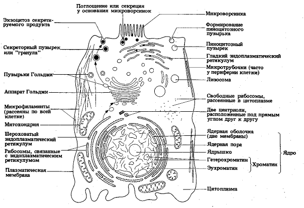
The structure of an animal cell. Cytoplasm of a living cell
Scientists position the animal cell as the main part of the body of a representative of the animal kingdom - both unicellular and multicellular.
They are eukaryotic, with a true nucleus and specialized structures - organelles that perform differentiated functions.
Plants, fungi and protists have eukaryotic cells; bacteria and archaea have simpler prokaryotic cells.
Structure animal cell different from plant. An animal cell does not have walls or chloroplasts (organelles that perform).
Drawing of an animal cell with captions
A cell consists of many specialized organelles that perform various functions.

Most often, it contains the majority, sometimes all existing types organelles
Basic organelles and organelles of an animal cell
Organelles and organelles are the “organs” responsible for the functioning of a microorganism.
Core
The nucleus is the source of deoxyribonucleic acid (DNA) - genetic material. DNA is the source of the creation of proteins that control the state of the body. In the nucleus, strands of DNA wrap tightly around highly specialized proteins (histones) to form chromosomes.

The nucleus selects genes to control the activity and functioning of the tissue unit. Depending on the type of cell, it contains various set genes. DNA is found in the nucleoid region of the nucleus where ribosomes are formed. The nucleus is surrounded by a nuclear membrane (karyolemma), a double lipid bilayer that separates it from the other components.
The nucleus regulates cell growth and division. When chromosomes are formed in the nucleus, they are duplicated during the process of reproduction, forming two daughter units. Organelles called centrosomes help organize DNA during division. The core is usually represented in the singular.
Ribosomes
Ribosomes are the site of protein synthesis. They are found in all tissue units, in plants and animals. In the nucleus, the DNA sequence that codes for a specific protein is copied into a free messenger RNA (mRNA) strand.
The mRNA strand travels to the ribosome via messenger RNA (tRNA), and its sequence is used to determine the arrangement of amino acids in the chain that makes up the protein. In animal tissue, ribosomes are located freely in the cytoplasm or attached to the membranes of the endoplasmic reticulum.
Endoplasmic reticulum
The endoplasmic reticulum (ER) is a network of membranous sacs (cisternae) extending from the outer nuclear membrane. It modifies and transports proteins created by ribosomes.

There are two types of endoplasmic reticulum:
- granular;
- agranular.
The granular ER contains attached ribosomes. The agranular ER is free of attached ribosomes and is involved in the creation of lipids and steroid hormones and the removal of toxic substances.
Vesicles
Vesicles are small spheres of lipid bilayer that are part of the outer membrane. They are used to transport molecules throughout the cell from one organelle to another and participate in metabolism.
Specialized vesicles called lysosomes contain enzymes that digest large molecules (carbohydrates, lipids and proteins) into smaller ones to facilitate their use by the tissue.
Golgi apparatus
The Golgi apparatus (Golgi complex, Golgi body) also consists of cisterns that are not interconnected (unlike the endoplasmic reticulum).

The Golgi apparatus receives proteins, sorts them, and packages them into vesicles.
Mitochondria
The process of cellular respiration occurs in mitochondria. Sugars and fats are broken down and energy is released in the form of adenosine triphosphate (ATP). ATP controls all cellular processes, mitochondria produce ATP cells. Mitochondria are sometimes called "generators".
Cell cytoplasm
Cytoplasm is the fluid environment of the cell. It can function even without a core, however, for a short time.
Cytosol
Cytosol is called cellular fluid. The cytosol and all the organelles within it, excluding the nucleus, are collectively called the cytoplasm. The cytosol is primarily composed of water and also contains ions (potassium, proteins, and small molecules).
Cytoskeleton
The cytoskeleton is a network of filaments and tubes distributed throughout the cytoplasm.

It performs the following functions:
- gives shape;
- provides strength;
- stabilizes tissue;
- secures organelles in certain places;
- plays an important role in signal transmission.
There are three types of cytoskeletal filaments: microfilaments, microtubules and intermediate filaments. Microfilaments are the smallest elements of the cytoskeleton, and microtubules are the largest.
Cell membrane
The cell membrane completely surrounds the animal cell, which does not have a cell wall, unlike plants. The cell membrane is a double layer consisting of phospholipids.

Phospholipids are molecules containing phosphates attached to glycerol and fatty acid radicals. They spontaneously form double membranes in water due to their simultaneously hydrophilic and hydrophobic properties.
The cell membrane is selectively permeable—it is capable of allowing certain molecules to pass through. Oxygen and carbon dioxide pass easily, while large or charged molecules must pass through a special channel in the membrane to maintain homeostasis.
Lysosomes
Lysosomes are organelles that degrade substances. The lysosome contains about 40 digestive enzymes. It is interesting that the cellular organism itself is protected from degradation in the event of a breakthrough of lysosomal enzymes into the cytoplasm; mitochondria that have completed their functions are subject to decomposition. After cleavage, residual bodies are formed, primary lysosomes turn into secondary ones.
Centriole
Centrioles are dense bodies located near the nucleus. The number of centrioles varies, most often there are two. The centrioles are connected by an endoplasmic bridge.
What does an animal cell look like under a microscope?
Under a standard optical microscope, the main components are visible. Due to the fact that they are connected into a constantly changing organism that is in motion, it can be difficult to identify individual organelles.

The following parts are not in doubt:
- core;
- cytoplasm;
- cell membrane.
A higher resolution microscope, a carefully prepared specimen, and some practice will help you study the cell in more detail.
Centriole functions
The exact functions of the centriole remain unknown. There is a widespread hypothesis that centrioles are involved in the division process, forming the division spindle and determining its direction, but there is no certainty in the scientific world.
The structure of a human cell - drawing with captions
A unit of human cell tissue has complex structure. The figure shows the main structures.

Each component has its own purpose; only in a conglomerate do they ensure the functioning of an important part of a living organism.
Signs of a living cell
A living cell is similar in its characteristics to a living being as a whole. It breathes, eats, develops, divides, and in its structure there are various processes. It is clear that the fading of natural processes for the body means death.
Distinctive features of plant and animal cells in the table
Plant and animal cells have both similarities and differences, which are briefly described in the table:
| Sign | Vegetable | Animal |
| Getting food | Autotrophic. Photosynthesizes nutrients |
Heterotrophic. Does not produce organic matter. |
| Power storage | In vacuole | In the cytoplasm |
| Storage carbohydrate | starch | glycogen |
| Reproductive system | Formation of a septum in the maternal unit | Formation of constriction in the maternal unit |
| Cell center and centrioles | In lower plants | All types |
| Cell wall | Dense, retains its shape | Flexible, allows change |
The main components are similar for both plant and animal particles.
Conclusion
An animal cell is a complex functioning organism with distinctive features, functions, purpose of existence. All organelles and organoids contribute to the life process of this microorganism.
Some components have been studied by scientists, while the functions and features of others have yet to be discovered.
Unlike eukaryotic and fungal cells, animal cells do not have. This feature was lost in the distant past by single-celled organisms that gave rise to. Most cells, both animal and plant, range in size from 1 to 100 µm (micrometers) and are therefore only visible with a microscope.
The earliest fossil evidence of animals dates from the Vendian period (650-454 million years ago). The first ended with this period, but during the subsequent period, an explosion of new life forms gave rise to many of the major faunal groups known today. There is evidence that animals appeared before the early (505-438 million years ago).
The structure of animal cells
Animal cell structure diagram
- - self-replicating organelles consisting of nine bundles of microtubules and found only in animal cells. They help organize cell division, but are not essential for this process.
- - necessary for cell movement. In multicellular organisms, cilia function to move fluid or substances around a stationary cell, or for or groups of cells.
- - a network of pouches that produces, processes and transports chemical compounds inside and outside the cell. It is associated with a two-layer nuclear envelope, providing a pipeline between the core and.
- Endosomes are membrane-bound vesicles formed by a complex set of processes known as endosomes, and are found in the cytoplasm of almost any animal cell. The basic mechanism of endocytosis is the opposite of what occurs during or cellular secretion.
- - distribution and delivery department chemical substances cells. It modifies proteins and fats embedded in the endoplasmic reticulum and also prepares them for export outside the cell.
- Intermediate filaments are a broad class of fibrous proteins that play important roles as both structural and functional elements.
Cytoplasm is called the internal environment of the body because it is constantly moving and moves all cellular components. The cytoplasm constantly undergoes metabolic processes and contains all organic and non-organic substances.
Structure
Cytoplasm consists of a permanent liquid part - hyaloplasm and elements that change - organelles and inclusions.
Organelles of the cytoplasm are divided into membrane and non-membrane, the latter in turn can be double-membrane and single-membrane.
- Non-membrane organelles: ribosomes, vacuoles, centrosome, flagella.
- Double membrane organelles: mitochondria, plastids, nucleus.
- Single-membrane organelles: Golgi apparatus, lysosomes, vacuoles, endoplasmic reticulum.
Also, the components of the cytoplasm include cellular inclusions, presented in the form of lipid droplets or glycogen granules.
The main features of the cytoplasm:
- Colorless;
- elastic;
- mucous-viscous;
- structured;
- movable.
The liquid part of the cytoplasm in its own way chemical composition differs in cells of different specializations. The main substance is water from 70% to 90%; it also contains proteins, carbohydrates, phospholipids, trace elements, and salts.
The acid-base balance is maintained at 7.1–8.5pH (slightly alkaline).
Cytoplasm, when studied at high magnification of a microscope, is not a homogeneous medium. There are two parts - one is located on the periphery in the area of the plasmalemma (ectoplasm), the other is near the core (endoplasm).
Ectoplasm serves as a link with the environment, intercellular fluid and neighboring cells. Endoplasm- This is the location of all organelles.
The structure of the cytoplasm contains special elements - microtubules and microfilaments.
Microtubules– non-membrane organelles necessary for the movement of organelles within the cell and the formation of the cytoskeleton. The globular protein tubulin is the main building block for microtubules. One tubulin molecule does not exceed 5 nm in diameter. In this case, the molecules are able to combine with each other, together forming a chain. 13 such chains form a microtubule with a diameter of 25 nm.
Tubulin molecules are found in constant movement for the formation of microtubules, if the cell is exposed to unfavorable factors, the process is disrupted. Microtubules are shortened or completely denatured. These elements of the cytoplasm are very important in the life of plants and bacterial cells, since they take part in the structure of their shells.

Microfilaments- These are submicroscopic non-membrane organelles that form the cytoskeleton. They are also part of the contractile apparatus of the cell. Microfilaments consist of two types of proteins - actin and myosin. Actin fibers are thin up to 5 nm in diameter, and myosin fibers are thick – up to 25 nm. Microfilaments are mainly concentrated in the ectoplasm. There are also specific filaments that are characteristic of a particular cell type.
Microtubules and microfilaments together form the cell cytoskeleton, which ensures the interconnection of all organelles and intracellular metabolism.
High molecular weight biopolymers are also isolated in the cytoplasm. They are combined into membrane complexes that permeate the entire internal space of the cell, determine the location of organelles, and delimit the cytoplasm from the cell wall.
The structural features of the cytoplasm lie in the ability to change its internal environment. It can exist in two states: semi-liquid ( sol) and viscous ( gel). So, depending on the influence external factors(temperature, radiation, chemical solutions), the cytoplasm passes from one state to another.
Functions
- Fills the intracellular space;
- connects all structural elements of the cell with each other;
- transports synthesized substances between organelles and outside the cell;
- establishes the location of organelles;
- is a medium for physical and chemical reactions;
- responsible for cell turgor, the constancy of the internal environment of the cell.
The functions of the cytoplasm in a cell also depend on the type of cell itself: plant, animal, eukaryotic or prokaryotic. But in all living cells, an important physiological phenomenon occurs in the cytoplasm - glycolysis. The process of glucose oxidation, which occurs under aerobic conditions and ends with the release of energy.
Movement of the cytoplasm
The cytoplasm is in constant motion; this characteristic is of great importance in the life of the cell. Thanks to movement, metabolic processes inside the cell and the distribution of synthesized elements between organelles are possible.
Biologists have observed the movement of cytoplasm in large cells, while monitoring the movement of vacuoles. Microfilaments and microtubules, which are activated in the presence of ATP molecules, are responsible for the movement of the cytoplasm.
The movement of the cytoplasm shows how active the cells are and how capable they are of survival. This process is dependent on external influences, so the slightest changes in environmental factors stop or accelerate it.
The role of the cytoplasm in protein biosynthesis. Protein biosynthesis is carried out with the participation of ribosomes, which are located directly in the cytoplasm or on the granular ER. Also, through nuclear pores, mRNA enters the cytoplasm, which carries information copied from DNA. The exoplasm contains the necessary amino acids for protein synthesis and enzymes that catalyze these reactions.
Summary table of the structure and functions of the cytoplasm
| Structural elements | Structure | Functions |
|---|---|---|
| Ectoplasm | Dense layer of cytoplasm | Provides connection with the external environment |
| Endoplasm | More fluid layer of cytoplasm | Location of cell organelles |
| Microtubules | Constructed from a globular protein - tubulin with a diameter of 5 nm, which is capable of polymerization | Responsible for intracellular transport |
| Microfilaments | Composed of actin and myosin fibers | Form the cytoskeleton, maintain connections between all organelles |
We invite you to familiarize yourself with the materials and.
: cellulose membrane, membrane, cytoplasm with organelles, nucleus, vacuoles with cell sap.Presence of plastids - main feature plant cell.
Functions of the cell membrane- determines the shape of the cell, protects against factors external environment.
Plasma membrane- a thin film, consists of interacting molecules of lipids and proteins, delimits the internal contents from the external environment, ensures the transport of water, minerals and organic matter by osmosis and active transfer, and also removes waste products.
Cytoplasm- the internal semi-liquid environment of the cell, in which the nucleus and organelles are located, provides connections between them, and participates in basic life processes.
Endoplasmic reticulum- a network of branching channels in the cytoplasm. It is involved in the synthesis of proteins, lipids and carbohydrates, and in the transport of substances. Ribosomes are bodies located on the ER or in the cytoplasm, consisting of RNA and protein, and are involved in protein synthesis. EPS and ribosomes are a single apparatus for the synthesis and transport of proteins.
Mitochondria- organelles delimited from the cytoplasm by two membranes. Organic substances are oxidized in them and ATP molecules are synthesized with the participation of enzymes. Increase in the surface of the inner membrane on which enzymes are located due to cristae. ATP is an energy-rich organic substance.
Plastids(chloroplasts, leucoplasts, chromoplasts), their content in the cell is the main feature of the plant organism. Chloroplasts are plastids containing the green pigment chlorophyll, which absorbs light energy and uses it to synthesize organic substances from carbon dioxide and water. Chloroplasts are separated from the cytoplasm by two membranes, numerous outgrowths - grana on the inner membrane, in which chlorophyll molecules and enzymes are located.
Golgi complex- a system of cavities delimited from the cytoplasm by a membrane. The accumulation of proteins, fats and carbohydrates in them. Carrying out the synthesis of fats and carbohydrates on membranes.
Lysosomes- bodies delimited from the cytoplasm by a single membrane. The enzymes they contain accelerate the breakdown of complex molecules into simple ones: proteins into amino acids, complex carbohydrates to simple ones, lipids to glycerol and fatty acids, and also destroy dead parts of the cell, whole cells.
Vacuoles- cavities in the cytoplasm filled with cell sap, a place of accumulation of reserve nutrients and harmful substances; they regulate the water content in the cell.
Core - main part cells covered on the outside with a two-membrane, pore-pierced nuclear envelope. Substances enter the core and are removed from it through the pores. Chromosomes are carriers of hereditary information about the characteristics of an organism, the main structures of the nucleus, each of which consists of one DNA molecule combined with proteins. The nucleus is the place of synthesis of DNA, mRNA, r-RNA.
The presence of an outer membrane, cytoplasm with organelles, and a nucleus with chromosomes.
Outer or plasma membrane- delimits the contents of the cell from environment(other cells, intercellular substance), consists of lipid and protein molecules, provides communication between cells, transport of substances into the cell (pinocytosis, phagocytosis) and out of the cell.
Cytoplasm- the internal semi-liquid environment of the cell, which provides communication between the nucleus and organelles located in it. The main life processes take place in the cytoplasm.
Cell organelles:
1) endoplasmic reticulum(EPS)- a system of branching tubules, participates in the synthesis of proteins, lipids and carbohydrates, in the transport of substances in the cell;
2) ribosomes- bodies containing rRNA are located on the ER and in the cytoplasm and participate in protein synthesis. EPS and ribosomes are a single apparatus for protein synthesis and transport;
3) mitochondria- “power stations” of the cell, delimited from the cytoplasm by two membranes. The inner one forms cristae (folds), increasing its surface. Enzymes on the cristae accelerate the oxidation of organic substances and the synthesis of energy-rich ATP molecules;
4) Golgi complex- a group of cavities delimited by a membrane from the cytoplasm, filled with proteins, fats and carbohydrates, which are either used in vital processes or removed from the cell. The membranes of the complex carry out the synthesis of fats and carbohydrates;
5) lysosomes- bodies filled with enzymes accelerate the breakdown of proteins into amino acids, lipids into glycerol and fatty acids, polysaccharides into monosaccharides. In lysosomes, dead parts of the cell, whole cells, are destroyed.
Cellular inclusions- accumulations of reserve nutrients: proteins, fats and carbohydrates.
Core- the most important part of the cell. It is covered with a double-membrane shell with pores, through which some substances penetrate into the nucleus, and others enter the cytoplasm. Chromosomes are the main structures of the nucleus, carriers of hereditary information about the characteristics of the organism. It is transmitted during the division of the mother cell daughter cells, and with germ cells - to daughter organisms. The nucleus is the site of DNA, mRNA, and rRNA synthesis.
Exercise:
Explain why organelles are called specialized cell structures?
Answer: Organelles are called specialized cell structures because they perform strictly certain functions, is stored in the kernel hereditary information, ATP is synthesized in mitochondria, photosynthesis occurs in chloroplasts, etc.
If you have questions about cytology, you can contact
Cytoplasm- this is limited cell membrane internal environment cells except the nucleus and vacuole. It was previously said that the cell consists of 80% water. A feature of the structure of the cell cytoplasm is that most of the cell’s water structure is in the cytoplasm. The solid part of the cytoplasm includes proteins, carbohydrates, phospholipids, cholesterol and other nitrogen-containing substances. organic compounds, mineral salts, inclusions in the form of glycogen droplets (in animal cells) and other substances. Almost all processes of cellular metabolism take place in the cytoplasm. The cytoplasm also contains reserve nutrients and insoluble waste products from metabolic processes.
Functions of the cytoplasm or the role of the cytoplasm in the cell
Functions of the cytoplasm or role of the cytoplasm:
1. Connect all parts of the cell into a single whole;
2. Chemical processes take place in it;
3. Transports substances;
4. Performs a support function.
TO structural features of the cytoplasm the following can be attributed:
1. Colorless viscous substance;
2. Is in constant motion;
3. Contains organoids (permanent structural components both cellular inclusions and non-permanent structural cells);
4. Inclusions can be in the form of drops (fats) and grains (proteins and carbohydrates).
You can see what the cytoplasm looks like using the example of the structure of a plant cell or animal cell.
Movement of the cytoplasm
The movement of cytoplasm in the cell is virtually continuous. The movement of the cytoplasm itself is carried out due to the cytoskeleton, or more precisely due to changes in the shape of the cytoskeleton.
Cytoplasmic organoids
Organoids of the cell cytoplasm include all organoids located in the cell, since they are all located inside the cytoplasm. All organelles in the cytoplasm are in a mobile state and can move due to the cytoskeleton.
Composition of the cytoplasm
The composition of the cytoplasm includes:
1. Water approximately 80%;
2. Protein about 10%;
3. Lipids about 2%;
4. Organic salts about 1%;
5. Inorganic salts 1%;
6. RNA approximately 0.7%;
7. DNA approximately 0.4%.
The above composition of the cytoplasm is true for eukaryotic cells.
Similar articles
 The best amulets against the evil eye and damage Amulet against the evil eye with hands for children
 How to read the Psalter correctly
 Delicious dishes with sausages
 A glimpse of Bella. Romantic chronicle. A glimpse of genius. Messerer about Akhmadulina Boris Messerer glimpse of Bella romantic chronicle
 I dreamed that I was sailing on a boat on the river
 How to cook beef entrecote in a frying pan
 About the company Foreign language courses at Moscow State University
 Which city and why became the main one in Ancient Mesopotamia?
 Why Bukhsoft Online is better than a regular accounting program!
 Which year is a leap year and how to calculate it