
Hydra reproduction: description, features. Hydra - class Hydrozoa: sensory organs, nervous and digestive systems, reproduction Hydra plant
The hydra's body looks like an oblong sac, the walls of which consist of two layers of cells - ectoderm And endoderm.
Between them lies a thin gelatinous non-cellular layer - mesoglea, serving as a support.
The ectoderm forms the covering of the animal’s body and consists of several types of cells: epithelial-muscular, intermediate And stinging.
The most numerous of them are epithelial-muscular.

Ectoderm
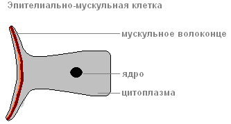
epithelial muscle cell
Due to muscle fibers, lying at the base of each cell, the body of the hydra can contract, lengthen and bend.
Between the epithelial-muscle cells there are groups of small, round cells with large nuclei and a small amount of cytoplasm, called intermediate.
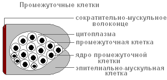
When the hydra's body is damaged, they begin to grow and divide rapidly. They can transform into other types of cells in the hydra body, except for epithelial-muscular ones.
The ectoderm contains stinging cells, serving for attack and defense. They are mainly located on the tentacles of the hydra. Each stinging cell contains an oval capsule in which the stinging filament is coiled.

Structure of a stinging cell with a coiled stinging thread
If prey or an enemy touches a sensitive hair located outside the stinging cell, in response to irritation the stinging thread is ejected and pierces the body of the victim.
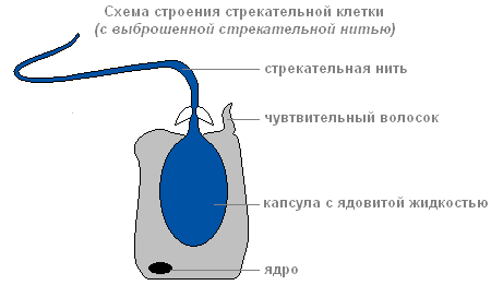
Structure of a stinging cell with discarded stinging thread
Through the thread channel, a substance that can paralyze the victim enters the victim’s body.
There are several types of stinging cells. The threads of some pierce the skin of animals and introduce poison into their bodies. The threads of others are wrapped around the prey. The threads of the third are very sticky and stick to the victim. Usually the hydra “shoots” several stinging cells. After the shot, the stinging cell dies. New stinging cells are formed from intermediate.
The structure of the inner layer of cells
Endoderm lines the entire intestinal cavity from the inside. It includes digestive-muscular And glandular cells.

Endoderm
Digestive system
There are more digestive muscle cells than others. Muscle fibers they are capable of reduction. When they shorten, the hydra's body becomes thinner. Complex movements (movement by “tumbling”) occur due to contractions of muscle fibers of ectoderm and endoderm cells.
Each of the digestive-muscle cells of the endoderm has 1-3 flagella. Hesitating flagella create a current of water, which drives food particles towards the cells. Digestive-muscle cells of the endoderm are capable of forming pseudopods, capture and digest small food particles in the digestive vacuoles.

The structure of the digestive muscle cell
Glandular cells in the endoderm secrete digestive juice into the intestinal cavity, which liquefies and partially digests food.
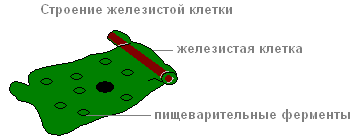
The structure of the glandular cell
Prey is captured by the tentacles using stinging cells, the venom of which quickly paralyzes small victims. By coordinated movements of the tentacles, the prey is brought to the mouth, and then, with the help of body contractions, the hydra is “put on” the victim. Digestion begins in the intestinal cavity ( cavity digestion), ends inside the digestive vacuoles of epithelial-muscular endoderm cells ( intracellular digestion). Nutrients are distributed throughout the hydra's body.
When the digestive cavity contains remains of the prey that cannot be digested, and waste from cellular metabolism, it contracts and empties.
Breath
Hydra breathes oxygen dissolved in water. She has no respiratory organs, and she absorbs oxygen over the entire surface of her body.
Circulatory system
Absent.
Selection
The release of carbon dioxide and other unnecessary substances formed during life processes is carried out from the cells of the outer layer directly into the water, and from the cells of the inner layer into the intestinal cavity, then out.
Nervous system
Below the skin-muscle cells are star-shaped cells. These are nerve cells (1). They connect with each other and form a nerve network (2).
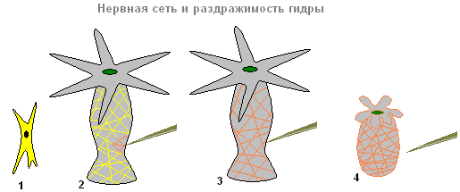
Nervous system and irritability of the hydra
If you touch the hydra (2), then excitation (electrical impulses) occurs in the nerve cells, which instantly spreads throughout the entire nervous network (3) and causes contraction of the skin-muscle cells and the entire body of the hydra shortens (4). The response of the hydra body to such irritation is unconditioned reflex.
Sex cells
With the approach of cold weather in the fall, germ cells are formed from intermediate cells in the ectoderm of the hydra.
There are two types of germ cells: eggs, or female germ cells, and sperm, or male germ cells.
The eggs are located closer to the base of the hydra, sperm develop in tubercles located closer to the mouth.
egg cell Hydra is similar to an amoeba. It is equipped with pseudopods and grows rapidly, absorbing neighboring intermediate cells.
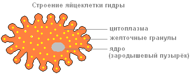
The structure of the hydra egg cell

The structure of the hydra sperm
Sperm in appearance they resemble flagellated protozoa. They leave the hydra's body and swim using a long flagellum.
Fertilization. Reproduction
The sperm swims up to the hydra with the egg cell and penetrates inside it, and the nuclei of both sex cells merge. After this, the pseudopods are retracted, the cell is rounded, a thick shell is released on its surface - an egg is formed. When the hydra dies and is destroyed, the egg remains alive and falls to the bottom. With the onset of warm weather, the living cell located inside the protective shell begins to divide, the resulting cells are arranged in two layers. From them a small hydra develops, which comes out through a break in the egg shell. Thus, the multicellular animal hydra at the beginning of its life consists of only one cell - an egg. This suggests that the ancestors of Hydra were single-celled animals.

Asexual reproduction of hydra
Under favorable conditions, hydra reproduces asexually. A bud forms on the animal’s body (usually in the lower third of the body), it grows, then tentacles form and a mouth breaks through. The young hydra buds from the mother's body (in this case, the mother and daughter polyps are attached with tentacles to the substrate and pull in different directions) and leads an independent lifestyle. In autumn, hydra begins to reproduce sexually. On the body, in the ectoderm, gonads are formed - sex glands, and in them, germ cells develop from intermediate cells. When hydra gonads form, a medusoid nodule is formed. This suggests that the hydra gonads are highly simplified sporifers, the last stage in the series of transformation of the lost medusoid generation into an organ. Most species of hydra are dioecious; hermaphroditism is less common. Hydra eggs grow rapidly by phagocytosis of surrounding cells. Mature eggs reach a diameter of 0.5-1 mm. Fertilization occurs in the body of the hydra: through a special hole in the gonad, the sperm penetrates the egg and merges with it. The zygote undergoes complete uniform fragmentation, as a result of which a coeloblastula is formed. Then, as a result of mixed delamination (a combination of immigration and delamination), gastrulation occurs. A dense protective shell (embryotheca) with spine-like outgrowths is formed around the embryo. At the gastrula stage, the embryos enter suspended animation. Adult hydras die, and the embryos sink to the bottom and overwinter. In the spring, development continues, in the parenchyma of the endoderm, an intestinal cavity is formed by divergence of cells, then the rudiments of tentacles are formed, and a young hydra emerges from under the shell. Thus, unlike most marine hydroids, hydra does not have free-swimming larvae and its development is direct.
Regeneration
Hydra has a very high ability to regenerate. When cut crosswise into several parts, each part restores the “head” and “leg”, maintaining the original polarity - the mouth and tentacles develop on the side that was closer to the oral end of the body, and the stalk and sole develop on the aboral side of the fragment. The whole organism can be restored from individual small pieces of the body (less than 1/100 of the volume), from pieces of tentacles, and also from a suspension of cells. Moreover, the regeneration process itself is not accompanied by increased cell division and is a typical example of morphallaxis.
Movement
In a calm state, the tentacles extend several centimeters. The animal slowly moves them from side to side, lying in wait for prey. If necessary, the hydra can move slowly.
"Walking" mode of transportation
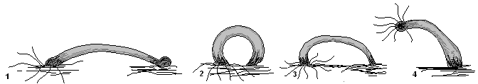
"Walking" method of movement of the hydra
Having curved its body (1) and attached its tentacles to the surface of an object (substrate), the hydra pulls the sole (2) to the front end of the body. Then the walking movement of the hydra is repeated (3,4).
"Tumbling" mode of movement

"Tumbling" method of movement of the hydra
In another case, it seems to tumble over its head, alternately attaching itself to objects with its tentacles and its sole (1-5).
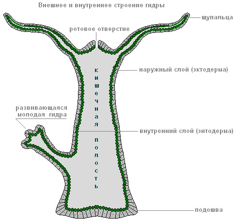
Figure: Structure of freshwater hydra. Radial symmetry of Hydra
Habitat, structural features and vital functions of the freshwater hydra polyp
In lakes, rivers or ponds with clean, transparent water, a small translucent animal is found on the stems of aquatic plants - polyp hydra(“polyp” means “multi-legged”). This is an attached or sedentary coelenterate animal with numerous tentacles. The body of an ordinary hydra has an almost regular cylindrical shape. At one end is mouth, surrounded by a corolla of 5-12 thin long tentacles, the other end is elongated in the form of a stalk with sole at the end. Using the sole, the hydra is attached to various underwater objects. The body of the hydra, together with the stalk, is usually up to 7 mm long, but the tentacles can extend several centimeters.
Radial symmetry of Hydra
If you draw an imaginary axis along the body of the hydra, then its tentacles will diverge from this axis in all directions, like rays from a light source. Hanging down from some aquatic plant, the hydra constantly sways and slowly moves its tentacles, lying in wait for prey. Since the prey can appear from any direction, the tentacles arranged in a radial manner best suit this method of hunting.
Radiation symmetry is characteristic, as a rule, of animals leading an attached lifestyle.
Hydra intestinal cavity
The body of the hydra has the form of a sac, the walls of which consist of two layers of cells - the outer (ectoderm) and the inner (endoderm). Inside the body of the hydra there is intestinal cavity(hence the name of the type - coelenterates).
The outer layer of hydra cells is the ectoderm.

Figure: structure of the outer layer of cells - hydra ectoderm
The outer layer of hydra cells is called - ectoderm. Under a microscope, several types of cells are visible in the outer layer of the hydra - the ectoderm. Most of all here are skin-muscular. By touching their sides, these cells create the cover of the hydra. At the base of each such cell there is a contractile muscle fiber, which plays an important role in the movement of the animal. When everyone's fiber skin-muscular cells contract, the hydra's body contracts. If the fibers contract on only one side of the body, then the hydra bends in that direction. Thanks to the work of muscle fibers, the hydra can slowly move from place to place, alternately “stepping” with its sole and tentacles. This movement can be compared to a slow somersault over your head.
The outer layer contains and nerve cells. They have a star-shaped shape, as they are equipped with long processes.
The processes of neighboring nerve cells come into contact with each other and form nerve plexus, covering the entire body of the hydra. Some of the processes approach the skin-muscle cells.
Hydra irritability and reflexes
Hydra is able to sense touch, temperature changes, the appearance of various dissolved substances in water and other irritations. This causes her nerve cells to become excited. If you touch the hydra with a thin needle, then the excitement from irritation of one of the nerve cells is transmitted along the processes to other nerve cells, and from them to the skin-muscle cells. This causes muscle fibers to contract, and the hydra shrinks into a ball.

Picture: Hydra's irritability
In this example, we get acquainted with a complex phenomenon in the animal body - reflex. The reflex consists of three successive stages: perception of irritation, transfer of excitation from this irritation along the nerve cells and response body by any action. Due to the simplicity of the hydra's organization, its reflexes are very uniform. In the future we will become familiar with much more complex reflexes in more highly organized animals.
Hydra stinging cells

Pattern: hydra string or nettle cells
The entire body of the hydra and especially its tentacles are seated with a large number stinging, or nettles cells. Each of these cells has a complex structure. In addition to the cytoplasm and nucleus, it contains a bubble-like stinging capsule, inside which a thin tube is folded - stinging thread. Sticking out of the cage sensitive hair. As soon as a crustacean, small fish or other small animal touches a sensitive hair, the stinging thread quickly straightens, its end is thrown out and pierces the victim. Through a channel passing inside the thread, poison enters the body of the prey from the stinging capsule, causing the death of small animals. As a rule, many stinging cells are fired at once. Then the hydra uses its tentacles to pull the prey to its mouth and swallow it. The stinging cells also serve the hydra for protection. Fish and aquatic insects do not eat hydras, which burn their enemies. The poison from the capsules is reminiscent of nettle poison in its effect on the body of large animals.
The inner layer of cells is the hydra endoderm

Figure: structure of the inner layer of cells - hydra endoderm
Inner layer of cells - endoderm A. The cells of the inner layer - the endoderm - have contractile muscle fibers, but the main role of these cells is to digest food. They secrete digestive juice into the intestinal cavity, under the influence of which the hydra’s prey softens and breaks down into small particles. Some of the cells of the inner layer are equipped with several long flagella (as in flagellated protozoa). The flagella are in constant motion and sweep particles towards the cells. The cells of the inner layer are capable of releasing pseudopods (like those of an amoeba) and capturing food with them. Further digestion occurs inside the cell, in vacuoles (like in protozoa). Undigested food remains are thrown out through the mouth.
The hydra has no special respiratory organs; oxygen dissolved in water penetrates the hydra through the entire surface of its body.
Hydra regeneration
The outer layer of the hydra's body also contains very small round cells with large nuclei. These cells are called intermediate. They play a very important role in the life of the hydra. With any damage to the body, intermediate cells located near the wounds begin to grow rapidly. From them, skin-muscle, nerve and other cells are formed, and the wounded area quickly heals.
If you cut a hydra crosswise, tentacles grow on one of its halves and a mouth appears, and a stalk appears on the other. You get two hydras.
The process of restoring lost or damaged body parts is called regeneration. Hydra has a highly developed ability to regenerate.
Regeneration, to one degree or another, is also characteristic of other animals and humans. Thus, in earthworms it is possible to regenerate a whole organism from their parts; in amphibians (frogs, newts) entire limbs, various parts of the eye, tail and internal organs can be restored. When a person is cut, the skin is restored.
Hydra reproduction
Asexual reproduction of hydra by budding

Figure: Hydra asexual reproduction by budding
Hydra reproduces asexually and sexually. In summer, a small tubercle appears on the hydra's body - a protrusion of the wall of its body. This tubercle grows and stretches out. Tentacles appear at its end, and a mouth breaks out between them. This is how the young hydra develops, which at first remains connected to the mother with the help of a stalk. Outwardly, all this resembles the development of a plant shoot from a bud (hence the name of this phenomenon - budding). When the little hydra grows up, it separates from the mother’s body and begins to live independently.
Hydra sexual reproduction
By autumn, with the onset of unfavorable conditions, hydras die, but before that, sex cells develop in their body. There are two types of germ cells: ovoid, or female, and spermatozoa, or male reproductive cells. Sperm are similar to flagellated protozoa. They leave the hydra's body and swim using a long flagellum.

Figure: Hydra sexual reproduction
The hydra egg cell is similar to an amoeba and has pseudopods. The sperm swims up to the hydra with the egg cell and penetrates inside it, and the nuclei of both sex cells merge. Happening fertilization. After this, the pseudopods are retracted, the cell is rounded, and a thick shell is formed on its surface - a egg. At the end of autumn, the hydra dies, but the egg remains alive and falls to the bottom. In the spring, the fertilized egg begins to divide, the resulting cells are arranged in two layers. From them a small hydra develops, which, with the onset of warm weather, comes out through a break in the egg shell.
Thus, the multicellular animal hydra at the beginning of its life consists of one cell - an egg.
From this article you will learn everything about the structure of freshwater hydra, its lifestyle, nutrition, and reproduction.
External structure of the hydra
Polyp (meaning "multipede") hydra is a tiny translucent creature that lives in the clean, transparent waters of slow-flowing rivers, lakes, and ponds. This coelenterate animal leads a sedentary or sedentary lifestyle. The external structure of freshwater hydra is very simple. The body has an almost regular cylindrical shape. At one of its ends there is a mouth, which is surrounded by a crown of many long thin tentacles (from five to twelve). At the other end of the body there is a sole, with the help of which the animal is able to attach to various objects under water. The body length of freshwater hydra is up to 7 mm, but the tentacles can greatly stretch and reach a length of several centimeters.
Radiation symmetry
Let's take a closer look at the external structure of the hydra. The table will help you remember their purpose.
The body of the hydra, like many other animals leading an attached lifestyle, is characterized by What is it? If you imagine a hydra and draw an imaginary axis along its body, then the animal’s tentacles will diverge from the axis in all directions, like the rays of the sun.

The structure of the hydra's body is dictated by its lifestyle. It attaches itself to an underwater object with its sole, hangs down and begins to sway, exploring the surrounding space with the help of tentacles. The animal is hunting. Since the hydra lies in wait for prey, which can appear from any direction, the symmetrical radial arrangement of the tentacles is optimal.
Intestinal cavity
Let's look at the internal structure of the hydra in more detail. The hydra's body looks like an oblong sac. Its walls consist of two layers of cells, between which there is an intercellular substance (mesoglea). Thus, there is an intestinal (gastric) cavity inside the body. Food enters it through the mouth opening. It is interesting that the hydra, which is not currently eating, has practically no mouth. The ectoderm cells close and grow together in the same way as on the rest of the body surface. Therefore, every time before eating, the hydra has to break through its mouth again.
The structure of freshwater hydra allows it to change its place of residence. There is a narrow opening on the sole of the animal - the aboral pore. Through it, liquid and a small bubble of gas can be released from the intestinal cavity. With the help of this mechanism, the hydra is able to detach from the substrate and float to the surface of the water. In this simple way, with the help of currents, it spreads throughout the reservoir.

Ectoderm
The internal structure of the hydra is represented by ectoderm and endoderm. The ectoderm is called the body-forming hydra. If you look at an animal under a microscope, you can see that the ectoderm includes several types of cells: stinging, intermediate and epithelial-muscular.
The most numerous group is skin-muscle cells. They touch each other with their sides and form the surface of the animal’s body. Each such cell has a base - a contractile muscle fiber. This mechanism provides the ability to move.
When all fibers contract, the animal’s body contracts, lengthens, and bends. And if the contraction occurs on only one side of the body, then the hydra bends. Thanks to this work of cells, the animal can move in two ways - “tumbling” and “stepping”.
Also in the outer layer are star-shaped nerve cells. They have long processes, with the help of which they come into contact with each other, forming a single network - a nerve plexus that entwines the entire body of the hydra. Nerve cells also connect with skin and muscle cells.
Between the epithelial-muscle cells there are groups of small, round-shaped intermediate cells with large nuclei and a small amount of cytoplasm. If the hydra's body is damaged, the intermediate cells begin to grow and divide. They can turn into anything

Stinging cells
The structure of hydra cells is very interesting; the stinging (nettle) cells with which the entire body of the animal, especially the tentacles, are strewn deserve special mention. have a complex structure. In addition to the nucleus and cytoplasm, the cell contains a bubble-shaped stinging chamber, inside which there is a thin stinging thread rolled into a tube.
A sensitive hair emerges from the cell. If prey or an enemy touches this hair, the stinging thread sharply straightens and is thrown out. The sharp tip pierces the victim’s body, and poison flows through the channel running inside the thread, which can kill a small animal.
Typically, many stinging cells are triggered. The hydra grabs prey with its tentacles, pulls it to its mouth and swallows it. The poison secreted by the stinging cells also serves for protection. Larger predators do not touch the painfully stinging hydras. The venom of the hydra is similar in effect to the poison of nettles.
Stinging cells can also be divided into several types. Some threads inject poison, others wrap around the victim, and others stick to it. After triggering, the stinging cell dies, and a new one is formed from the intermediate one.

Endoderm
The structure of hydra also implies the presence of such a structure as the inner layer of cells, endoderm. These cells also have muscle contractile fibers. Their main purpose is to digest food. Endoderm cells secrete digestive juices directly into the intestinal cavity. Under its influence, the prey is split into particles. Some endoderm cells have long flagella that are constantly in motion. Their role is to pull food particles towards the cells, which in turn release pseudopods and capture food.
Digestion continues inside the cell and is therefore called intracellular. Food is processed in vacuoles, and undigested remains are thrown out through the mouth. Breathing and excretion occurs through the entire surface of the body. Let us consider once again the cellular structure of the hydra. The table will help you do this clearly.
Reflexes
The structure of the hydra is such that it is able to sense changes in temperature, the chemical composition of water, as well as touch and other stimuli. The nerve cells of an animal are capable of being excited. For example, if you touch it with the tip of a needle, the signal from the nerve cells that sensed the touch will be transmitted to the rest, and from the nerve cells to the epithelial-muscular cells. The skin-muscle cells will react and contract, the hydra will shrink into a ball.

Such a reaction is bright. It is a complex phenomenon consisting of successive stages - perception of the stimulus, transmission of excitation and response. The structure of the hydra is very simple, therefore the reflexes are monotonous.
Regeneration
The cellular structure of the hydra allows this tiny animal to regenerate. As mentioned above, intermediate cells located on the surface of the body can transform into any other type.
With any damage to the body, the intermediate cells begin to divide, grow very quickly and replace the missing parts. The wound is healing. The regenerative abilities of the hydra are so high that if you cut it in half, one part will grow new tentacles and a mouth, and the other will grow a stem and sole.

Asexual reproduction
Hydra can reproduce both asexually and sexually. Under favorable conditions in the summer, a small tubercle appears on the animal’s body and the wall protrudes. Over time, the tubercle grows and stretches. Tentacles appear at its end and a mouth breaks through.
Thus, a young hydra appears, connected to the mother’s body by a stalk. This process is called budding because it is similar to the development of a new shoot in plants. When a young hydra is ready to live on its own, it buds off. The daughter and mother organisms attach to the substrate with tentacles and stretch in different directions until they separate.
Sexual reproduction
When it starts to get colder and unfavorable conditions are created, the turn of sexual reproduction begins. In the fall, hydras begin to form sex cells, male and female, from the intermediate ones, that is, egg cells and sperm. The egg cells of hydras are similar to amoebas. They are large, strewn with pseudopods. Sperm are similar to the simplest flagellates; they are able to swim with the help of a flagellum and leave the body of the hydra.
After the sperm penetrates the egg cell, their nuclei fuse and fertilization occurs. The pseudopods of the fertilized egg retract, it becomes rounded, and the shell becomes thicker. An egg is formed.
All hydras die in the fall, with the onset of cold weather. The mother's body disintegrates, but the egg remains alive and overwinters. In the spring it begins to actively divide, the cells are arranged in two layers. With the onset of warm weather, the small hydra breaks through the shell of the egg and begins an independent life.
The body shape of the hydra is tubular. The mouth opening of these animals is covered with tentacles. Hydras live in water, and with their stinging tentacles they kill and bring prey to their mouths.
Type - CoelenteratesClass - Hydroid
Genus/Species - Hydra vulgaris, H.oligactis, etc.
Basic data:
DIMENSIONS
Length: 6-15 mm.
REPRODUCTION
Vegetative: has a budding character. A bud appears on the body of the mother, from which the daughter gradually develops.
Sexual: Most species of hydra are dioecious. The gonads contain cells from which eggs develop. Sperm cells develop in the testis.
LIFESTYLE
Habits: live in fresh and brackish waters.
Food: plankton, fish fry, ciliates.
Lifespan: no data.
RELATED SPECIES
The phylum Coelenterata includes more than 9,000 species, some of them (15-20) live only in fresh waters.
Freshwater hydras are one of the smallest predators. Despite this, they are able to provide themselves with food. Hydras have a tubular body shape. Using their soles, they attach themselves to underwater plants or rocks and move their tentacles in search of prey. Green hydras contain photosynthetic algae.
FOOD
 Hydra is a predatory animal that lives in water. It feeds on small organisms living in water, for example, ciliates, oligochaete worms, planktonic crustaceans, water fleas, insects and their larvae, and fish fry. A hydra that hunts attaches itself to an aquatic plant, branch or leaf and hangs on it. Her tentacles are very wide open. They constantly make circular searching movements. If one of them touches the victim, others rush towards it. Hydra paralyzes prey with stinging cell venom. The hydra uses its tentacles to pull its paralyzed prey towards its mouth. She swallows small animals whole. If the prey is larger than the hydra, the predator opens its mouth wide and the walls of its body stretch. If such prey is so large that it does not fit into the gastric cavity, then the hydra swallows only part of it and, to the extent of digestion, pushes the victim deeper and deeper.
LIFESTYLE
 Hydras live alone. However, in places that are especially rich in food, several hydras hunt at once. This happens because the water current brings a lot of food to a certain place. Hydras of the Nuiga genus prefer fresh water. These animals were discovered by the researcher who invented the microscope, A. Leeuwenhoek (1632-1723). Another scientist, G. Tremblay, discovered that hydras easily restore lost body parts. An inconspicuous tubular body, crowned with tentacles that grow around the mouth opening, and a sole at the end of the body are the main features of the hydra's appearance. The gastric cavity of this animal is continuous. The tentacles are hollow. The body walls consist of two layers of cells. There are glandular cells located in the middle part of the hydra's body. The different types are very similar to each other. They differ mainly in color (and, as a result, different colors indicate some structural feature). Bright green hydras have symbiotic algae living in their bodies. Hydras react to light and swim towards it. These animals are sedentary. They spend most of their lives in an attached state, waiting for prey. With the sole, like a suction cup, hydras are firmly attached to plants.
 REPRODUCTION
 Hydras reproduce in two ways - sexual and vegetative. Vegetative propagation is represented by budding. Under suitable external conditions, several buds develop on the hydra’s body. At the very beginning, the bud looks like a small mound, later miniature tentacles appear at its outer end. The tentacles grow and stinging cells appear on them. The lower part of the body of the daughter individual becomes thinner, the hydra's mouth opens, the young individual branches off and begins an independent life. These animals reproduce by budding in the warm season. With the onset of autumn, hydras begin sexual reproduction. Sex cells are formed in the gonads. The gonad cracks and an egg emerges. Around the same time, sperm are formed in the testes of other hydras. They also leave the gonad and swim in the water. One of them fertilizes the egg. An embryo develops in the egg. Protected by a double shell, it overwinters at the bottom. In the spring, a fully formed hydra emerges from the egg.
 DID YOU KNOW THAT...
- Hydra does not age because every cell in its body is renewed within a few weeks. This animal lives only in the warm season. With the beginning of winter, all adult hydras die. Only their eggs, protected by a strong double shell - the embryotheca, can survive the winter.
- Hydras easily restore their lost limbs. The scientist G. Tremblay (1710-1784), as a result of his numerous experiments, obtained a seven-headed polyp, from which severed heads grew back. He looked like a mythical creature - the Lernaean Hydra, defeated by the hero of ancient Greece - Hercules.
- During constant movements in the water, the hydra performs quite original acrobatic tricks.
CHARACTERISTIC FEATURES OF HYDRA
Tentacles: the mouth opening is surrounded by a corolla with 5-12 tentacles with stinging cells. With their help, the animal paralyzes its prey and pulls it into its mouth. A hydra that hunts attaches itself to a hard surface and, spreading its tentacles widely, makes circular searching movements with them.Body: body shape is tubular. At the anterior end is a mouth opening surrounded by tentacles. The aboral pore is located in the middle of the sole. The hydra wall consists of two layers of cells. Digestive processes take place in the midsection of the body.
Mouth opening: covered with a corolla of tentacles. With its tentacles, the hydra pulls the animal into its mouth and swallows it.
Leg: The rear end of the hydra is narrowed - this is a leg that has a sole at the end.
Gonads: are formed in the ectoderm and have the appearance of tubercles. Sex cells accumulate in them.
Dome: length about 13 mm. This is for self-defense. The hydra rises and forms a dense dome.
Bud: The vegetative propagation of hydra has the nature of budding. Several buds may appear on the body at the same time. The buds are growing quickly.

PLACES OF ACCOMMODATION
Freshwater hydras live in fresh and brackish waters. They inhabit rivers, lakes, swamps and other bodies of water. The most common species are the common and brown hydra.
PRESERVATION
Each species of a genus living in a certain territory. These days they are not in danger of extinction.
To the class hydroid include invertebrate aquatic cnidarians. In their life cycle, two forms are often present, replacing each other: polyp and jellyfish. Hydroids can gather in colonies, but solitary individuals are also not uncommon. Traces of hydroids are found even in Precambrian layers, but due to the extreme fragility of their bodies, the search is very difficult.
A bright representative of hydroids - freshwater hydra, single polyp. Its body has a sole, a stalk and long tentacles relative to the stalk. She moves like a rhythmic gymnast - with each step she makes a bridge and somersaults over her “head”. Hydra is widely used in laboratory experiments; its ability to regenerate and high activity of stem cells, providing “eternal youth” to the polyp, prompted German scientists to search and study the “immortality gene.”
Hydra cell types
1. Epithelial-muscular cells form the outer covers, that is, they are the basis ectoderm. The function of these cells is to shorten the body of the hydra or make it longer; for this they have muscle fibers.
2. Digestive-muscular cells are located in endoderm. They are adapted to phagocytosis, capture and mix food particles that enter the gastric cavity, for which each cell is equipped with several flagella. In general, flagella and pseudopods help food penetrate from the intestinal cavity into the cytoplasm of hydra cells. Thus, her digestion occurs in two ways: intracavitary (for this there is a set of enzymes) and intracellular.
3. Stinging cells located primarily on the tentacles. They are multifunctional. Firstly, the hydra defends itself with their help - a fish that wants to eat the hydra is burned with poison and throws it away. Secondly, the hydra paralyzes prey captured by its tentacles. The stinging cell contains a capsule with a poisonous stinging thread; on the outside there is a sensitive hair, which, after irritation, gives a signal to “shoot”. The life of a stinging cell is short-lived: after being “shot” by a thread, it dies.
4. Nerve cells, together with shoots similar to stars, lie in ectoderm, under a layer of epithelial-muscle cells. Their greatest concentration is at the sole and tentacles. When exposed to any impact, the hydra reacts, which is an unconditional reflex. The polyp also has such a property as irritability. Let us also remember that the “umbrella” of a jellyfish is bordered by a cluster of nerve cells, and the body contains ganglia.
5. Glandular cells release a sticky substance. They are located in endoderm and promote food digestion.
6. Intermediate cells- round, very small and undifferentiated - lie in ectoderm. These stem cells divide endlessly, are capable of transforming into any other, somatic (except epithelial-muscular) or reproductive cells, and ensure the regeneration of the hydra. There are hydras that do not have intermediate cells (hence, stinging, nerve and reproductive cells), capable of asexual reproduction.
7. Sex cells develop into ectoderm. The egg cell of the freshwater hydra is equipped with pseudopods, with which it captures neighboring cells along with their nutrients. Among the hydras there is hermaphroditism, when eggs and sperm are formed in the same individual, but at different times.
Other features of freshwater hydra
1. Hydras do not have a respiratory system; they breathe over the entire surface of the body.
2. The circulatory system is not formed.
3. Hydras eat larvae of aquatic insects, various small invertebrates, and crustaceans (daphnia, cyclops). Undigested food remains, like other coelenterates, are removed back through the mouth.
4. Hydra is capable of regeneration, for which intermediate cells are responsible. Even when cut into fragments, the hydra completes the necessary organs and turns into several new individuals.
Similar articles
 Delicious dishes with sausages
 A glimpse of Bella. Romantic chronicle. A glimpse of genius. Messerer about Akhmadulina Boris Messerer glimpse of Bella romantic chronicle
 I dreamed that I was sailing on a boat on the river
 How to cook beef entrecote in a frying pan
 About the company Foreign language courses at Moscow State University
 Which city and why became the main one in Ancient Mesopotamia?
 Why Bukhsoft Online is better than a regular accounting program!
 Which year is a leap year and how to calculate it
 Prayer for lighting a lamp at home
 What was their strength and what was their weakness?